




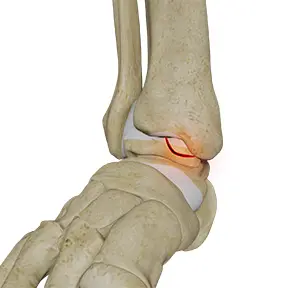
Subtalar distraction bone-block arthrodesis is a mouthful, but the operation itself is straightforward to describe: fusing the joint just below the ankle, with a piece of bone graft inserted to maintain the heel’s height. It’s commonly used to treat the malunited heel-bone fracture — one that healed with the foot a bit shortened and the heel out of alignment.
The skin problem
The textbook approach to the subtalar joint comes in from the lateral side — the outside of the foot. The skin there is thin and prone to breaking down, especially when there has been a previous calcaneal fracture, previous open surgery, prior infection, or significant scarring. Performing the operation through compromised skin is a recipe for wound breakdown and deep infection. So in those cases, the operation has historically been off-limits.
What we did
This 2021 paper, on which I was second author, describes a medial approach to the subtalar joint — coming in from the inside of the foot, where the skin envelope is often more forgiving. The paper details the key surgical landmarks, the safe planes of dissection, and the technical considerations for getting good visualisation of the joint from this less-familiar side.
Why it matters
Surgical approaches matter. Two surgeons can perform the same fusion through completely different access routes, and the choice between them is often the difference between a smooth recovery and a wound-related disaster. Having a validated medial approach in the toolkit means we can offer this fusion to patients who would otherwise have been turned down because the lateral skin wasn’t safe.
What this means for patients
If you need a subtalar fusion but have a history of open injury, previous open surgery, or significant lateral foot scarring, the medial approach is a real alternative. Discuss it explicitly with your surgeon. The right side to come in from is the side where the skin is most likely to heal.
Source: Haldar A, Heidari N, Malagelada F. Medial approach for subtalar distraction bone block arthrodesis Foot (Edinburgh) 2021;49:101848. PMID: 34583132. Read the full paper on PubMed →