Osteochondral Injuries
An osteochondral injury (OCL) is damage to the cartilage and the bone underneath — most often on the talus, after a significant ankle sprain or twist that didn't fully heal.
An osteochondral injury (OCL) is damage to the cartilage and the bone underneath — most often on the talus, after a significant ankle sprain or twist that didn't fully heal.
Foot & Ankle · Professor Nima Heidari
An osteochondral injury (OCL) is damage to the cartilage and the bone underneath — most often on the talus, after a significant ankle sprain or twist that didn't fully heal.
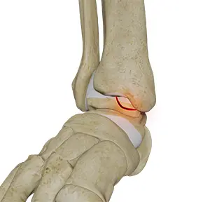
An osteochondral lesion (OCL) of the talus is an area of damage to the cartilage and the bone immediately beneath it on the dome of the talus — the lower bone of the ankle joint. The cartilage normally provides a smooth, low-friction surface; when it is injured, the joint no longer glides as it should.
Most lesions follow an injury, very often an ankle sprain or fracture, where the talus is compressed against the tibia. Some appear suddenly after a single significant injury; others develop gradually and are only found when ankle pain fails to settle. They are a common and frequently missed reason for an ankle that keeps hurting long after a sprain should have recovered.
Typical symptoms are deep, poorly localised ankle pain that is worse with activity, intermittent swelling, and sometimes catching, clicking or a sense of the ankle giving way. Because the lesion sits inside the joint, plain X-rays often look normal — an MRI or CT is usually needed to show the true size of the defect and whether the underlying bone is involved.
Smaller, stable lesions often settle with a period of protected weight-bearing, activity modification and physiotherapy. When symptoms persist, keyhole (arthroscopic) surgery allows the lesion to be assessed directly and treated — by removing unstable fragments and stimulating a healing response, or, for larger defects, by techniques that restore the cartilage and bone such as grafting or biological repair.
The right treatment depends on the size and depth of the lesion, how long it has been present, and your activity demands. The priority is always to preserve the joint and return you to full, confident weight-bearing.
An ankle that won’t settle six months after a sprain isn’t a slow ligament — it is often an osteochondral lesion that wasn’t looked for.
Professor Heidari
Clinical approach
MRI maps the size, location, and bone involvement. CT is added when planning a graft procedure.
Small lesions: arthroscopic debridement, sometimes with microfracture or AMIC. Larger or deeper lesions: OATS or MACI. The decision is anatomical, not preferential.
OCLs are rarely isolated — ligamentous instability is often present. Addressing both at the same operation gives the cartilage repair the best chance.
Treatments considered
Immobilising and not allowing weight to be placed on the joint, followed by a course of physiotherapy can suffice in allowing the bone and cartilage to heal. Should this not be effective or in the case of serious injury, your consultant may recommend arthroscopic surgery to take away the injured cartilage and bone and remodel the bone’s surface to reduce pain and cut the risk of arthritis developing.
Common questions
A consultation is a structured conversation, not a sales pitch. The diary holds while you decide.
Book a Consultation →Signed off on 26 August 2026