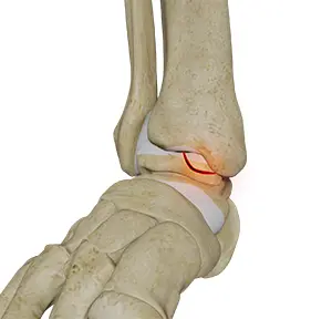





For decades, the imaging a surgeon relies on during an operation has been 2D fluoroscopy — flat X-ray pictures, one plane at a time. It is fast and familiar. But foot and ankle fractures often involve curved joint surfaces, and a small misalignment can hide between those flat views — only to show up later on a CT scan after the patient has woken up. Sometimes that means a second trip to theatre.
Bringing the CT into the room
The O-arm is a 3D scanner that can be brought into the operating theatre, giving the surgeon a CT-quality, multi-plane picture while the patient is still asleep and the wound is still open. This study, which Professor Heidari co-authored, describes two foot-and-ankle trauma cases in detail.
What we found
In both cases, the 3D scan revealed a problem the standard 2D images had missed — either the fracture had not been fully reduced, or an implant’s position needed adjusting. Crucially, because it was caught during the operation, the surgeon could revise the fixation there and then. Both patients went on to heal.
What this means for patients
The goal of fracture surgery is to get it right the first time — a well-aligned joint heals better and is far less likely to wear out early. Intra-operative 3D imaging is one of the ways surgeons close the gap between “looked fine on the X-ray” and “was actually perfect on the CT”, sparing patients the second operation that gap sometimes costs.
Source: Jeyaseelan L, Malagelada F, Parker L, Panagopoulos A, Heidari N, Vris A. Intra-Operative 3-Dimensional Imaging (O-arm) in Foot and Ankle Trauma Surgery: Report of 2 Cases and Review of the Literature. The Open Orthopaedics Journal. Read the full paper →