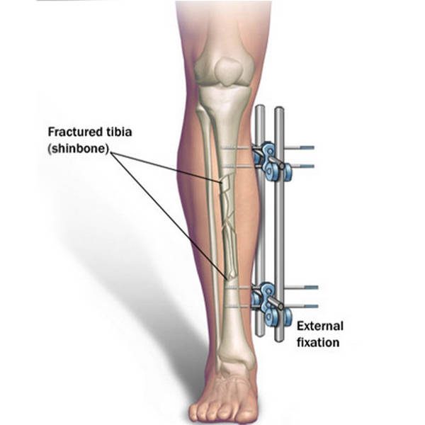





One of the underappreciated complications of fixing a tibial fracture with an internal nail is rotational malalignment — the bone heals, but the leg ends up subtly twisted compared to the other side. Walking, running, ankle-loading patterns — all subtly off. The reported incidence is up to 20%.
The measurement problem
Detecting rotational malalignment depends entirely on the accuracy of measurement. Historically, surgeons have done this in three ways: by clinical examination, by plain X-rays, or by CT scans where the operator manually measures axes on different slices. All three depend on the operator’s eye and are inherently subjective. Two clinicians measuring the same leg will produce different numbers.
What we did
This 2021 paper, on which I was a co-author, introduced a fully digital technique using CT-based motion analysis (CTMA) software. The principle: take CT scans of both legs, build 3D reconstructions, mathematically merge the proximal portions of the two legs and let the software calculate exactly how much the distal portions are out of alignment. The operator’s eye is removed from the calculation.
What we found
Across 10 pairs of cadaver lower legs we confirmed that healthy lower legs are symmetrical in the axial and coronal planes — the assumption all rotation-measurement techniques implicitly rely on. We also identified a small sagittal-plane difference between the left and right sides, which would benefit from further study with a larger sample size before being interpreted clinically.
What this means for patients
If you are several months post-tibial-nail and walking with a subtle but persistent gait abnormality, ask whether your surgeon can use a modern digital CT analysis to measure rotation, rather than estimating from the films. The newer technique gives an objective number that the older methods can’t match.
Source: Bakhshayesh P, Ihediwa U, Sandher S, Vris A, Heidari N, Enocson A. A novel technique to assess rotational deformities in lower extremities using CT-based motion analysis Scientific Reports 2021;11(1):21123. PMID: 34702869. Read the full paper on PubMed →