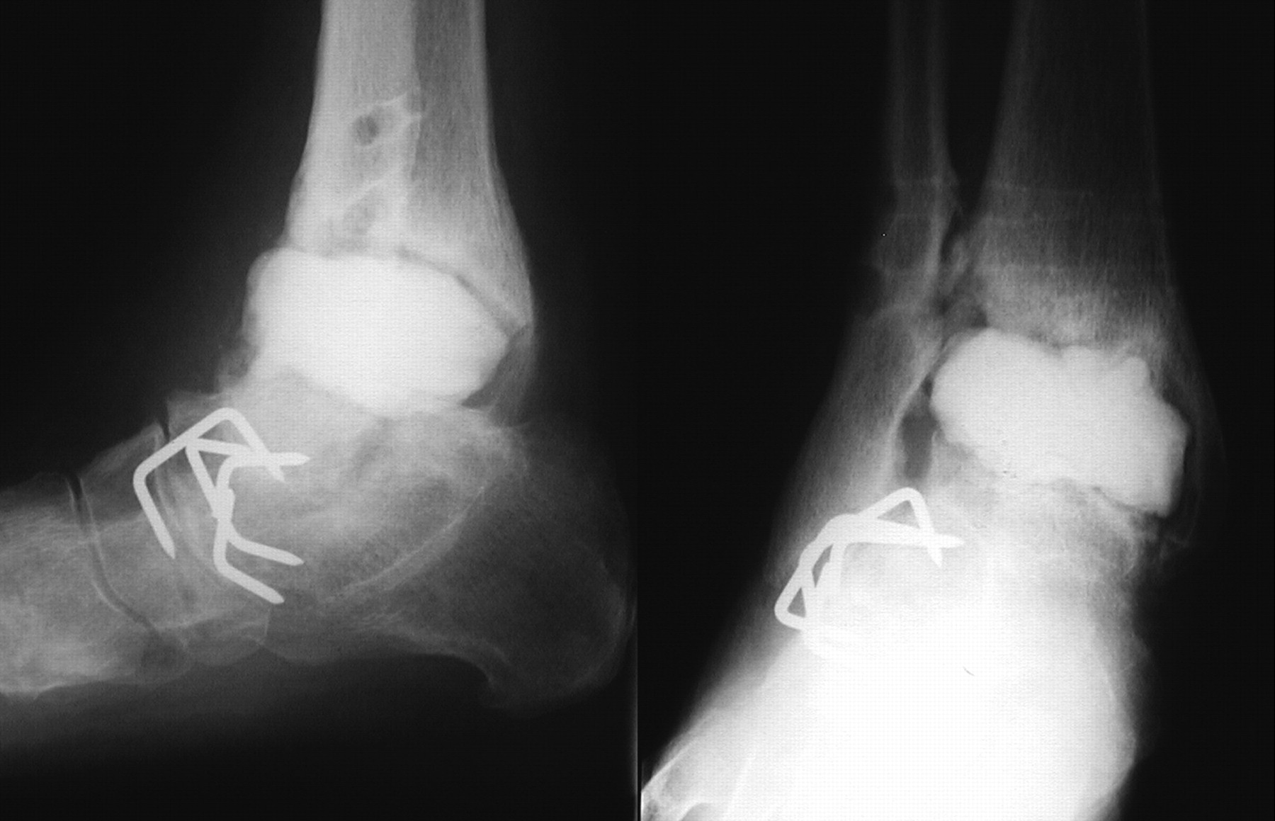





One of the most consequential decisions in diabetic foot infection is the choice of antibiotic. Get it right and the foot heals, the limb is saved. Get it wrong — even by something as subtle as targeting the wrong species of bacteria — and the infection grinds on, the bone keeps eroding, and we end up months later having a much harder conversation about reconstruction or amputation.
Why surface samples aren’t enough
For decades the default was to take a swab of the wound surface and send it to the lab. The problem is that diabetic foot wounds are colonised — the skin around the ulcer is teeming with bacteria that aren’t actually causing the deep infection. A surface swab tells you what’s on top, not what’s in the bone.
What a bone biopsy does
A bone biopsy — a small sample of bone taken under sterile conditions, usually as a brief procedure under local or short general anaesthetic — gives us the actual organism living in the bone. The lab can then identify it precisely and tell us exactly which antibiotic will kill it. There is also a second, useful effect: the biopsy itself removes a small amount of infected bone, contributing to the source-control side of treatment.
What we concluded in this 2019 paper
Together with my co-authors I argued that bone biopsies should be a routine part of working up diabetic foot osteomyelitis, not a special case reserved for when antibiotics have already failed. The earlier we have the right organism identified, the earlier we are on the right antibiotic, and the better the outcome.
What this means for you
If you’re a patient with a non-healing diabetic foot ulcer that has reached or threatens the bone, it is reasonable to ask your team whether a bone biopsy is part of the plan. It is the strongest piece of evidence we can use to choose your treatment.
Source: Heidari N, Kwok I, Vris A, Charalambous A. Should Treatment of Diabetic Foot Osteomyelitis Be Based on Bone Biopsies? Foot & Ankle International 2019. PMID: 31322951. Read the full paper on PubMed →